CT Scanner
The CT (computed tomography) scanner is a medical imaging technique that uses X-rays and computers to generate cross-sectional images (or "slices") of the internal structures of the body. It was invented in the 1970s by Godfrey Hounsfield and Allan Cormack, who shared the 1979 Nobel Prize in Medicine for their work.
The key innovations that enabled the development of the CT scanner were the linked technologies of X-ray generators, detectors, and computers. X-ray tubes and detectors had been in use for decades, but earlier scanners could only capture a single X-ray projection image from one angle. To generate a cross-sectional image, multiple projection images from different angles were required. This was not technologically feasible until the invention of minicomputers in the 1960s, which could rapidly process the large datasets needed for tomographic reconstruction.
Hounsfield realized that a focused beam of X-rays could be aimed at the patient from multiple angles around their body, and the resulting transmission measurements were processed by a computer to reconstruct an image of the slice. The first clinically useful CT scanner, the EMI Mark I, was installed in 1971. It collected data relatively slowly but demonstrated the viability of computed tomography. Within a few years, faster scanners were developed using improved detectors and minicomputer technology. This established CT scanning as a revolutionary medical imaging technique.
Modern multi-detector CT scanners can rapidly acquire large volumes of high-resolution data, enabling advanced 3D and 4D visualizations. But the groundwork was laid in the 1960s and 1970s by the convergence of x-ray imaging technology and the computational power of early minicomputers.

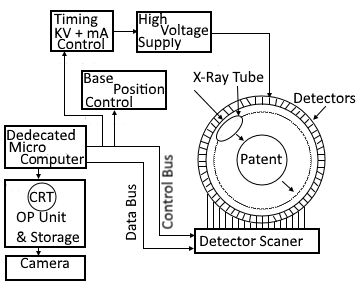
Operations of CT Scanner
- High voltage supply drives the X-ray tube that can be mechanically rotated along the circumference of a gantry.
- The patient is lying in a tube in the center of the gantry.
- The X-rays pass through the patient and produce an image on
detectors, which are fixed in a place around the circumference of the gantry in a large quantity. - The microcomputer senses the position of the tube in the gantry and samples
the output of the detector scanner which is opposite to the X-ray
tube. - A calculation based on the data of a computer scan of the tube is made by the computer.
- The output unit then produces a visual image of a transverse plane
across the section of the patient. - Output may be displayed on the cathode ray tube or photographed with a camera to produce a hard copy record.
Detector operation and construction
Detectors consist of ionization chambers filled with a gas such as xenon, sealed at both ends and having two conductors forming a capacitor on the sides. A high DC voltage is applied to the capacitors and x-rays entry. The chamber ionizes the xenon item causing it to migrate to the capacitor plate and causing a current in the high-voltage load. This current is proportional to the radiation and is fed to the computer as data for computing the image.
The advanced model of the CT scanner creates an image at an angle other than 90° by tilting the gantry.
Application of CT Scanner
- Diagnosing causes of pain - CT imaging can reveal problems in areas like the abdomen, pelvis, spine, head, chest, and extremities by detecting abnormal structures or sources of pain.
- Cancer screening and diagnosis - CT scans are used to detect tumors, determine their location and size, and assess their spread. They are commonly used for cancers of the lung, liver, kidney, and bowel.
- Detecting cardiovascular disease - CT angiography can visualize arteries to check for blockages, aneurysms, and buildup of plaque. Cardiac CT can assess coronary arteries and help diagnose heart conditions.
- Neurological disorders - CT can reveal internal brain abnormalities like tumors, bleeds, strokes, and skull fractures. It is often used in head trauma, stroke, and headache patients.
- Guiding medical procedures - CT is used to guide biopsies, radiation therapy, and minimally invasive surgery procedures by providing visualizations of the patient's anatomy.
- Bone imaging - CT excellently depicts fractures, orthopedic injuries, osteoarthritis, and bone tumors. 3D reconstructions clearly show complex fractures.
- Dental/sinus imaging - CT captures fine details of teeth and jaws. It is commonly used to assess dental implant sites and examine sinus anatomy.
- Emergency medicine - CT scanners are standard equipment in most emergency rooms, allowing rapid diagnosis of trauma, abdominal pain, and other conditions.
Side Effects and Risks of CT Scanner
- Radiation exposure - CT scans involve higher radiation doses compared to regular X-rays. There are increased lifetime risks of cancer, especially with multiple CT scans. However, the benefits usually outweigh the small risk.
- Allergic reactions - Some patients may experience allergic reactions to the intravenous contrast dye used in some CT scans. Mild itching and nausea are common, but severe reactions like difficulty breathing can occur.
- Kidney dysfunction - Contrast dyes can temporarily affect kidney function, especially in those with pre-existing kidney disease. Adequate hydration helps minimize this risk.
- Adverse effects on the fetus - CT scans are avoided during pregnancy unless absolutely necessary, as there are risks of birth defects or childhood cancer from excessive radiation.
- Claustrophobia - Some patients experience anxiety or claustrophobia due to the enclosed space of the CT scanner. Mild sedatives can help in such cases.
- False positives - CT scans may reveal incidental findings that are not clinically significant but cause patient anxiety and lead to additional testing.
- Hearing damage - Loud noise from the CT scanner rotating around the patient can damage hearing if protection isn't worn.
- Metal artifacts - Dental fillings or implants can distort images due to streaks or shadows. Artifacts interfere with obtaining optimal diagnostic images.
While adverse effects are rare when protocols are followed, doctors always weigh if the benefits of a CT scan outweigh any small risks for each patient. Proper usage and safety measures help make CT scanning a very low-risk procedure.
